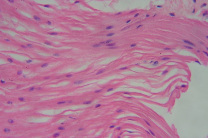
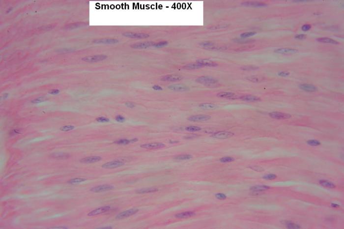
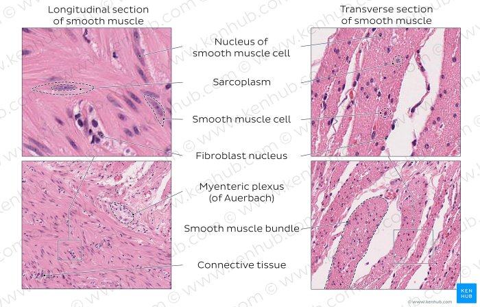
Smooth muscle under microscope 400x reveals a unique cellular structure that sets it apart from other muscle types. Its spindle-shaped morphology, central nucleus, and lack of striations are key identifying features that provide valuable insights into its function and pathology.
This detailed examination unveils the intricate mechanisms underlying smooth muscle’s role in various bodily systems, its response to hormonal and neurotransmitter regulation, and its involvement in common pathological conditions.
Smooth Muscle under Microscope 400x

Microscopic examination of smooth muscle at 400x magnification reveals several characteristic features.
Smooth Muscle Morphology
Smooth muscle cells are spindle-shaped with a central nucleus. They lack the striations that are characteristic of skeletal and cardiac muscle.
| Characteristic | Smooth Muscle | Skeletal Muscle | Cardiac Muscle |
|---|---|---|---|
| Shape | Spindle-shaped | Cylindrical | Branched |
| Nucleus | Central | Peripheral | Central |
| Striations | Absent | Present | Present |
Smooth Muscle Function
Smooth muscle is found in the walls of hollow organs, such as the digestive system, respiratory system, and blood vessels. It plays a role in regulating the flow of fluids and solids through these organs.
Smooth muscle contracts and relaxes through a process called the sliding filament mechanism. This mechanism involves the interaction of actin and myosin filaments, which slide past each other to shorten or lengthen the muscle fiber.
Smooth muscle function is regulated by hormones and neurotransmitters. For example, acetylcholine causes smooth muscle to contract, while epinephrine causes it to relax.
Smooth Muscle Histology
Smooth muscle tissue is prepared for microscopic examination by fixing it in a formalin solution and then embedding it in paraffin. The tissue is then cut into thin sections and stained with a hematoxylin and eosin (H&E) stain.
H&E staining reveals the characteristic spindle-shaped appearance of smooth muscle cells. The nucleus is typically located in the center of the cell and is surrounded by a thin rim of cytoplasm.
Smooth Muscle Pathology, Smooth muscle under microscope 400x
Common pathological conditions that affect smooth muscle include hyperplasia, hypertrophy, and neoplasia.
Hyperplasia is an increase in the number of smooth muscle cells. This can occur in response to a variety of stimuli, such as injury or inflammation.
Hypertrophy is an increase in the size of smooth muscle cells. This can occur in response to increased workload, such as in the case of hypertension.
Neoplasia is the formation of a tumor. Smooth muscle tumors can be benign or malignant.
Common Queries
What are the characteristic features of smooth muscle cells under 400x magnification?
Smooth muscle cells exhibit a spindle-shaped appearance, a centrally located nucleus, and a lack of striations, distinguishing them from skeletal and cardiac muscle cells.
How does smooth muscle function differ from skeletal and cardiac muscle?
Smooth muscle exhibits involuntary contractions and relaxes slowly compared to skeletal muscle, which contracts voluntarily and rapidly, and cardiac muscle, which contracts rhythmically and involuntarily.
What are the common pathological conditions that affect smooth muscle?
Smooth muscle can undergo hyperplasia (increased cell number), hypertrophy (increased cell size), and neoplasia (tumor formation), which can impact organ function and contribute to various diseases.

